Hydrocephalus
Diagnosis and surgical treatment of hydrocephalus and raised intracranial pressure.
- Specialty
- Brain
- Urgency
- Specialist evaluation
- Evaluation
- Imaging + examination
- Focus
- Hydrocephalus
- Dr. Nikhil Bhamare · Reviewed by
- 15+ years · Experience
- Medinova Super Speciality Hospital · Hospital
Overview
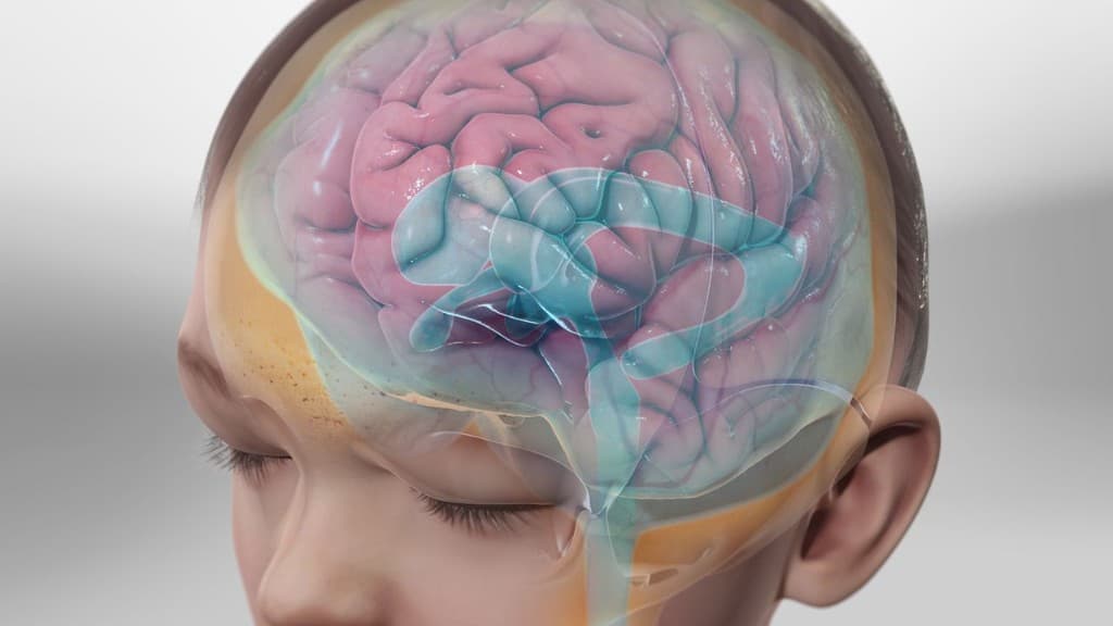
What is Hydrocephalus?
Diagnosis and surgical treatment of hydrocephalus and raised intracranial pressure.
Early recognition and accurate diagnosis are essential to prevent permanent neurological damage and guide the right treatment pathway — whether medical management, emergency surgery, or intensive care.
Dr. Nikhil Bhamare provides comprehensive evaluation using advanced diagnostic imaging and evidence-based treatment planning at Medinova Super Speciality Hospital, Nashik.
Recognise the signs
Symptoms
Understanding how hydrocephalus presents helps you seek evaluation before irreversible damage occurs.
Headache
Often described as sudden, severe, or the worst headache of life.
Vomiting
May occur with raised intracranial pressure or acute neurological injury.
Gait imbalance
Difficulty walking or loss of balance may reflect spinal cord or brain involvement.
Memory problems
Cognitive changes can reflect swelling, bleeding, or reduced brain perfusion.
In infants: increasing head size
A clinically significant symptom that should be evaluated by a neurosurgeon.
Clinical pathway
How diagnosis works
A structured evaluation from first symptoms to treatment decision — the standard of care in neurosurgery.
- Step 1
Symptoms
Recognition of sudden or progressive neurological warning signs.
- Step 2
Neurological examination
Focused assessment of strength, sensation, speech, and reflexes.
- Step 3
CT scan
Emergency imaging to detect bleeding, fracture, or mass effect.
- Step 4
MRI
Detailed evaluation when soft-tissue or spinal pathology is suspected.
- Step 5
Diagnosis
Correlation of imaging, examination, and clinical history.
- Step 6
Treatment decision
Medical, surgical, or ICU-based plan tailored to urgency and anatomy.
Causes & risk factors
Treatment options
Evidence-based management
Treatment is tailored to urgency, imaging findings, and neurological status — from emergency intervention to rehabilitation.
CT/MRI
Rapid non-invasive imaging to detect bleeding, swelling, or structural injury.
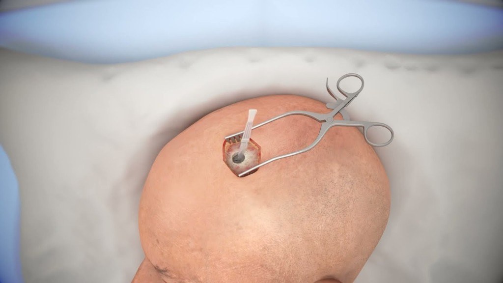
VP shunt
Surgical diversion of cerebrospinal fluid when normal drainage is impaired.
Endoscopic third ventriculostomy referral
Continuous monitoring of consciousness, blood pressure, and neurological status.
Long-term follow-up
Personalised neurosurgical evaluation and evidence-based management.
Recovery & rehabilitation
Long-term outcomes depend on early treatment, structured rehabilitation, and specialist follow-up.
- Neurological monitoring during the acute phase
- Physiotherapy and mobility rehabilitation
- Speech or occupational therapy when needed
- Gradual return to daily activities with specialist follow-up
Related procedures
Surgical and interventional options commonly used in the management of hydrocephalus.
Next step
Concerned about symptoms?
Early diagnosis can prevent permanent neurological damage. Dr. Nikhil Bhamare provides comprehensive evaluation for brain, spine, and nerve conditions using advanced diagnostic imaging and evidence-based treatment planning.
- · Urgent and elective consultations
- · Second opinions with imaging review
- · Emergency neurosurgical pathways at Medinova Super Speciality Hospital

Dr. Nikhil Bhamare
Neurosurgeon & Spine Surgeon
15+ years experience